Retinal diseases refer to a group of conditions that affect the retina—the thin, light-sensitive layer of tissue at the back of the eye responsible for detecting light and sending visual signals to the brain. Because the retina plays a central role in vision, changes or damage to this tissue may affect clarity, central vision, peripheral vision, or the ability to see in low light. Retinal conditions can arise from ageing, systemic illnesses, structural weaknesses, or changes in the blood vessels that supply the retina. Many retinal diseases develop gradually and may not cause early symptoms, while others may present more suddenly. Early diagnosis and timely management are important to help monitor retinal health over time.

Retinal diseases may be caused by a variety of underlying factors that affect retinal tissue, blood supply, or structural integrity. Some conditions involve deterioration of retinal cells, while others arise from fluid leakage, bleeding, or abnormal blood vessel growth. Common contributing causes include ageing, chronic medical conditions, genetic tendencies, and previous eye trauma or inflammation. Certain diseases such as diabetes or hypertension may affect the blood vessels of the retina, leading to swelling or leakage. High myopia can also increase the risk of retinal thinning or tears. In some cases, retinal changes develop without obvious symptoms, making regular examination important.

Retinal diseases progress at different rates depending on their type and underlying cause. In the early stages, individuals may have risk factors such as diabetes, high blood pressure, high myopia, or a family history of retinal disorders, yet experience no symptoms. As changes develop, subtle distortion, difficulty seeing in dim conditions, or areas of blurred vision may appear. Further progression may lead to fluid accumulation, bleeding, or damage to the central macula, affecting detailed vision needed for reading and recognising faces. In more serious disease, there may be substantial changes in vision if the retina is severely affected. Early evaluation and ongoing monitoring help identify risks and guide appropriate management.

Presence of risk factors
but no symptoms
Mild distortion, blurring,
or reduced contrast
Noticeable visual disturbances such as waviness, floaters, or dim spots
Central or peripheral vision loss depending on condition
Risk of significant or permanent vision loss if the retina becomes severely compromised
Retinal diseases may present with a wide range of symptoms depending on the area of the retina affected and the speed of progression. Some conditions affect central vision, while others impact peripheral sight or cause sudden visual disturbances. Because early symptoms may be subtle, regular eye examinations are important for at-risk individuals.


Age-Related Macular Degeneration (AMD) affects the macula, the small central area of the retina responsible for sharp, detailed vision used for reading, recognising faces, and seeing fine detail. In AMD, the cells of the macula gradually deteriorate with age, leading to distortion or loss of central vision while peripheral vision usually remains unaffected. In the early stages, waste deposits called drusen accumulate beneath the retina, interfering with the normal function of retinal cells. As the condition progresses, two different pathways may occur. In dry AMD, the macular cells slowly thin and lose function over time, causing gradual blurring of central vision. In wet AMD, fragile abnormal blood vessels grow beneath the retina and may leak fluid or bleed, causing sudden distortion, dark patches, or more rapid central vision loss. Although AMD does not cause total blindness, the central vision changes can affect daily activities such as reading, driving, and recognising faces. Early diagnosis and timely management play an important role in monitoring the condition and addressing changes as they arise, and in guiding ongoing care for central vision.

Diabetic Retinopathy occurs when long-term elevated blood sugar levels affect the tiny blood vessels that supply the retina—the light-sensitive layer at the back of the eye responsible for forming clear images. Over time, high glucose levels weaken these vessels, causing them to swell, leak fluid, or bleed. In the early stage, known as non-proliferative diabetic retinopathy, the vessel walls become fragile, leading to small retinal haemorrhages and fluid accumulation that can distort vision. When swelling affects the macula—the central area responsible for fine detail—this results in diabetic macular edema, a major cause of vision changes in diabetes. As the condition progresses, the retina becomes deprived of oxygen, triggering the growth of abnormal new blood vessels. This later stage, called proliferative diabetic retinopathy, is more severe and can lead to further complications. These fragile new vessels can bleed into the vitreous cavity, cause scar tissue to form, or pull on the retina, increasing the risk of retinal detachment. Without early detection and timely treatment, diabetic retinopathy can be associated with irreversible vision loss, but regular eye examinations and diabetes control may help lower the likelihood of more severe changes.

Retinal Vein Occlusion (RVO) occurs when one of the veins responsible for draining blood from the retina becomes blocked, similar to a “stroke” affecting the eye. The retina relies on healthy blood flow to function, and when a retinal vein is obstructed—often due to hardening of the vessel wall or pressure from a nearby artery—blood and fluid begin to back up within the retinal tissue. This congestion causes swelling, bleeding, and leakage of fluid into areas critical for vision. When the central retina (the macula) becomes swollen, a condition known as macular edema, vision may become blurry or distorted. Depending on where the blockage occurs, RVO may affect either the central retinal vein (central retinal vein occlusion) or one of its smaller branches (branch retinal vein occlusion). In some cases, the lack of oxygen in the retina triggers the growth of abnormal new blood vessels, which can bleed or lead to further complications such as glaucoma. The impact on vision can range from mild blurring to more marked loss, depending on the severity of the blockage and the extent of swelling or bleeding. Early detection and timely management are important in supporting ongoing care and monitoring retinal changes over time.
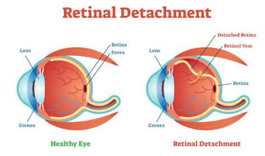
Retinal Tear and Retinal Detachment occur when the thin, light-sensitive tissue lining the back of the eye (the retina) becomes disrupted or pulled away from its normal position. A retinal tear often develops when the vitreous—the clear, gel-like substance that fills the centre of the eye—shrinks with age and tugs on the retina. If the traction is strong enough, it can create a small break or opening in the retinal tissue. Once a tear forms, fluid from inside the eye can seep through the opening and build up behind the retina. This accumulation of fluid can lift the retina off the underlying layers that supply it with oxygen and nutrients, leading to a retinal detachment.
When the retina detaches, the affected area can no longer function properly, resulting in shadows, flashes of light, or the sudden appearance of floaters. If the detachment spreads toward the central retina (the macula), central vision can become distorted or severely impaired. A retinal detachment is considered an urgent condition because prolonged separation can lead to permanent changes in vision. Early detection and prompt treatment of retinal tears may help reduce the likelihood of progression to a detachment, while timely surgical intervention is often required to reposition the retina and support its anatomical alignment.

Epiretinal Membrane (ERM) occurs when a thin layer of scar-like tissue forms on the surface of the macula—the central part of the retina responsible for sharp, detailed vision. This membrane develops from cells that migrate and grow on the retinal surface, often as a result of age-related changes in the vitreous, inflammation, retinal tears, previous eye surgery, or other retinal conditions. As the membrane contracts or tightens, it creates traction on the macula, causing the delicate retinal layers to wrinkle or distort. This distortion affects how light is focused onto the retina, leading to blurred or wavy central vision. Straight lines may appear crooked, fine detail becomes harder to see, and small print may seem unclear. In later situations, the membrane can thicken and pull more firmly on the macula, leading to greater visual distortion or reduced central vision clarity. Peripheral (side) vision usually remains unaffected. While ERM does not cause total blindness, it can affect daily tasks that require sharp, central vision, such as reading, sewing, or recognising faces. Early detection helps monitor progression, and in cases where the traction markedly affects vision, surgery may be recommended to remove the membrane and relieve the macular distortion.

Macular Hole occurs when a small full-thickness opening forms in the macula, the central part of the retina responsible for detailed, sharp vision. This condition usually develops as the vitreous—the clear gel that fills the centre of the eye—naturally shrinks and pulls away from the retina with age. In most people, the vitreous separates cleanly, but in some cases, it remains firmly attached to the macula. As it continues to tug on this delicate tissue, the persistent traction can stretch and eventually tear the macular layers, creating a hole. Once a hole forms, the central area of the retina cannot process light properly, leading to blurred, distorted, or missing central vision. Straight lines may appear bent, and reading or recognising faces may become challenging. In the early stages, the hole may be small and only mildly affect vision, but if the traction persists, it can gradually enlarge and cause more marked vision loss. Peripheral (side) vision typically remains unaffected, as the damage is limited to the macula. In rare cases, trauma, high myopia, or other retinal conditions can also contribute to macular hole formation. Prompt assessment and timely treatment—usually through a surgical procedure called vitrectomy—can help close the hole and address changes affecting central vision.

Summary & Comparison
| Retinal Condition | Symptoms | Causes |
|---|---|---|
|
Age-Related Macular Degeneration (AMD) |
• Blurred or distorted central vision |
• Age-related degeneration of macula |
|
Diabetic Retinopathy |
• Blurred or fluctuating vision |
• Long-term high blood sugar damaging retinal vessels |
|
Retinal Vein Occlusion (RVO) |
• Sudden blurred or distorted vision |
• Blockage of retinal vein by clot or vessel hardening |
|
Retinal Tear & Detachment |
• Sudden onset of new floaters |
• Vitreous pulling on retina as it shrinks (PVD) |
|
Epiretinal Membrane (ERM) |
• Wavy or distorted central vision |
• Cells growing on macular surface forming a membrane |
|
Macular Hole |
• Blurred or distorted central vision |
• Vitreous pulling strongly on macula |

Retinal tests are specialised examinations that allow eye specialists to assess the health of the retina, macula, and underlying blood vessels with detail. Because many retinal conditions develop quietly or cause subtle early symptoms, these tests play a role in identifying disease at earlier and more manageable stages. Current imaging technologies capture cross-sectional views of the retina, measure changes in thickness, identify fluid or bleeding, and map areas where vision may be affected. Other tests evaluate how well the retina functions by assessing visual fields, light sensitivity, or the clarity of the central macula. Together, these investigations help diagnose conditions such as macular degeneration, diabetic retinopathy, retinal vein occlusion, epiretinal membranes, and retinal tears or detachments. They also provide information for monitoring progression and guiding treatment decisions over time, so that any changes in retinal structure or function can be detected early and reviewed in a timely manner.
Retinal treatment focuses on addressing the underlying cause of disease, supporting vision, and managing changes to the delicate tissues at the back of the eye. Because retinal conditions can range from slow, progressive changes to sudden vision-threatening events, treatment is tailored to the specific diagnosis and its severity. Current retinal care combines imaging, targeted medications, laser therapy, and microsurgical techniques to manage a wide spectrum of conditions such as macular degeneration, diabetic retinopathy, retinal vein occlusion, epiretinal membranes, macular holes, and retinal detachments. Many conditions, especially those involving swelling or abnormal blood vessel growth, are managed with targeted injections that reduce abnormal vessel activity and leakage, manage retinal swelling, and address changes that may affect vision. Laser procedures may be used to seal weak areas, stop bleeding, or reduce the chance that retinal tears progress to detachment. In some situations, surgeries such as vitrectomy or scleral buckling may be required to repair the retina and reposition it. Early diagnosis and timely treatment are important, as retinal diseases can progress quietly yet be associated with irreversible vision loss if not addressed. Regular monitoring allows the retinal specialist to adjust treatment based on response and guide ongoing care.

Every patient’s condition, lifestyle, and goals are unique — and so should their care. We take the time to listen, understand, and evaluate your needs in detail. From there, we develop a tailored treatment plan that delivers the most ideal outcomes.

For most of us, our vision is our most valued sense. Whether you are a senior, of working age, or the parent of a child who needs help, we will prioritise understanding your case and determining the most suitable treatment to improve or preserve your vision. We welcome complex cases as we care about you, your sight and your future.

We harness the power of ophthalmic technology to deliver precise diagnostics, accurate treatments, and faster recovery times. From imaging systems to minimally invasive surgical tools, our investment in technology allows us to provide safe and effective eye care.
Your safety is our highest priority. Every diagnosis and treatment we provide is evidence-based to ensure that each step of your care is not only effective but also held to a high standard of safety and quality.

For pre-surgery, we will discuss all possible treatment options and recommends the most appropriate one for every patient. For post-surgery, we provide aftercare, including follow-up consultations and treatment plans tailored to every patient to promote optimal recovery.
Wish to book a consultation or have a question? WhatsApp Us!